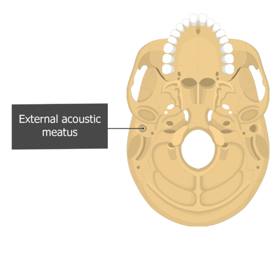


Sometimes there is a small contribution from the small auricular branch of the vagus nerve (CN X)įacial nerve (CN VII) innervates the external auricular muscles and probably the auricular skin as well Greater auricular nerve of the cervical plexus supplies the medial auricle and the lateral auricle posterior to the EACĪuriculotemporal nerve from the mandibular division of the trigeminal nerve (CN V3) supplies the skin of the lateral auricle anterior to the EAC The posterior auricular branch of the external carotid artery The tympanic membrane, is anatomically part of, and represents, the most medial extent of the external ear.Īn outer layer with a radial fiber arrangementĪn inner layer with a circular fiber arrangement It has an outer cover of extremely thin skin and an inner layer of cuboidal epithelium facing the tympanic cavity. The tympanic membrane (or tympanum) consists of two layers of collagen fibers: It is approximately 3 cm long and is lined by skin containing hair follicles (tragi), sebaceous glands, and ceruminous glands (which produce cerumen). The external auditory meatus is a short S-shaped canal within the tympanic temporal bone leading from the external acoustic pore of the auricle to the tympanic membrane. Intertragic incisure: a notch separating the tragus from the antitragusĬavum conchae: the deepest depression in the auricle, inferior to the crus of the helixĬymba conchae: depression surrounded by the crus of the helix below and the inferior crus of antihelix aboveĮar lobe ( lobule): the lowest part of the ear and the only part that does not contain cartilage, situated below the intertragic incisure Tragus: prominence in front of the external acoustic poreĬan be manually pushed back over the pore, to mitigate noiseĪntitragus: situated in the lower part of the antihelix and faces the tragus Scaphoid fossa / scapha: the depression between the helix and antihelix
#ACOUSTIC MEATUS FREE#
Helix: posterior free margin of the auricleĬrus helicis: anterior terminal portion of the helix superior to the external acoustic poreĬrura antihelicis: a pair of limbs located above the external acoustic poreįossa triangularis: tiny depression between the crura The auricle has a complex shape that is composed of several ridges, notches, and grooves (see Figure 1): It is composed of an irregular concave plate of elastic cartilage and dense connective tissue, covered by skin which contains short hairs (tragi), sebaceous glands, and ceruminous glands.

88, 608–617, Dec., 1968.The auricle is the part of the ear that projects laterally from the head. and Hurley, B.J.: Iophendylate Examination of Posterior Fossa in Diagnosis of Cerebellopontine Angle Tumours, Arch. Acoustic Neuroma: The adaptation of polytomography and lophendylate to the Early diagnosis of Acoustic Tumours. Merrill, Viruta: Atlas of Roentgenographic Positions: Vol. Saunders Company, Philadelphia & London, 2nd Edition, 1969. Shambaugh, Jr George E.: Surgery of the Ear, W. C.: Positioning in Radiography, London, Ilford Limited, Wm. & Whillis J.: Gray’s Anatomy, Longmans, Green & Co. E.: The Radiological Diagnosis of Acoustic Neuromas. K.: Radiology of Internal Auditory Canal, Indian Journal of Otolaryngology, Vol. and House, W.F.: X-Ray Diagnosis of Acoustic Neuromas, Arch. Zimmerman, H.M.: Panel on Tumours of Nervous System: Proceedings of National Cancer Conference, 1949, New York: American Cancer Society, pp.


 0 kommentar(er)
